


Chest pain that is new, persistent, worsening, or difficult to explain deserves more than symptom-based guesswork. You will learn when to call emergency services, which patterns need prompt assessment, what information to record, and what a first heart evaluation usually involves.
Key takeaways
- Call emergency services for chest pain lasting more than a few minutes.
- Treat pain that eases and returns as an emergency warning.
- Track timing, triggers, symptoms, and medical history before evaluation.
- Expect an examination and ECG before clinicians choose further tests.
When chest pain needs emergency help immediately
Call emergency services immediately for a chest pain emergency, not a routine appointment, if you have pressure, squeezing, fullness, heaviness, or severe pain lasting more than a few minutes. Call again without waiting if the pain eases and returns.
The answer to when to call emergency services for chest pain is also immediate action when pain comes with:
- Breathlessness, cold sweating, nausea, fainting, or light-headedness
- Pain spreading to an arm, shoulder, back, neck, jaw, or upper abdomen
While waiting for help:
- Stop exertion and sit down.
- Do not drive yourself to hospital.
- Unlock the door if you are alone.
- Follow the dispatcher’s instructions.
- Do not take aspirin automatically. Follow local emergency guidance, and tell the dispatcher about an aspirin allergy, bleeding risk, anticoagulant use, or any history of bleeding.
Heart attack warning signs do not always include crushing pain. Mild heart attack symptoms can begin gradually, come and go, or feel like pressure, breathlessness, unusual weakness, nausea, or back discomfort.
Women, older adults, and people with diabetes are more likely to have these less typical patterns, so wait for neither dramatic pain nor certainty before calling. A routine evaluation is appropriate only when emergency symptoms are absent and the pain has fully settled; new, persistent, worsening, or unexplained pain still needs prompt medical assessment.
How heart-related pain differs from common non-cardiac causes
Heart-related pain is more concerning when exertion brings on pressure, heaviness, squeezing, or fullness that improves with rest. Heart pain vs muscle pain is not always obvious, and heart symptoms can occur without dramatic pain.
| Pattern | More consistent with | Important clues |
|---|---|---|
| Exertional pressure or heaviness | Heart problem | Starts with walking or climbing and eases with rest; breathlessness, sweating, nausea, or pain spreading to the arm, jaw, back, or shoulder increases concern |
| Chest pain after eating | Acid reflux | Burning behind the breastbone, sour taste, belching, or worse pain when lying down |
| Pain after lifting, coughing, or an awkward movement | Muscle strain | Local soreness that increases when you move the torso or affected arm |
| Chest pain reproducible by pressing | Rib or chest-wall pain | Pressing one spot brings back the pain |
| Chest pain when breathing | Lung or chest-wall cause | Sharp pain with a deep breath, cough, or certain positions; infection can add fever or cough |
| Tightness during fear or a panic episode | Anxiety | Rapid breathing, trembling, tingling, or intense fear, after excluding physical causes |
These patterns guide triage; they do not diagnose the cause. Tenderness, positional pain, or pain linked to breathing makes a non-cardiac cause more likely but does not safely rule out a heart problem.
Seek urgent evaluation for new unexplained pain, severe or worsening pain, or symptoms in someone with diabetes, smoking history, high blood pressure, high cholesterol, kidney disease, obesity, prior vascular disease, or a strong family history of premature heart disease. Recurrent pain after a stent or bypass also needs reassessment.
Which chest-pain patterns and risk factors lower your threshold for evaluation
Recurring chest pain with exertion that predictably improves within minutes of rest is compatible with stable angina symptoms and needs medical assessment, even if the pain has stopped. New unexplained pain also deserves prompt assessment rather than self-diagnosis as indigestion or muscle strain.
| Pattern | What it suggests | What to do |
|---|---|---|
| Chest pain with exertion that eases with rest | A pattern compatible with stable angina | Arrange prompt medical assessment |
| Chest pain at rest | Possible acute coronary syndrome | Seek urgent medical assessment |
| More frequent or longer episodes | Possible worsening coronary blood flow | Seek urgent assessment |
| Pain triggered by less exertion than before | A change in the usual pattern | Seek urgent assessment |
Lower your threshold for evaluation if you have any of these cardiac risk factors:
- Known coronary artery disease
- A previous heart attack or stroke
- Diabetes
- High blood pressure
- High cholesterol
- Smoking
- Chronic kidney disease
- Obesity
- A strong family history of premature heart disease
These factors raise concern but do not prove that a particular episode is cardiac. Their absence is not reassuring: heart disease can occur without recognised risk factors. Recurrent pain after a stent or bypass also requires reassessment, because it can reflect progression elsewhere, in-stent narrowing, graft disease, or a non-cardiac cause.
If symptoms are worsening, persistent, or accompanied by breathlessness, sweating, nausea, faintness, or spreading pain, use emergency services rather than waiting for a routine appointment.
What to record and expect at a first heart evaluation
Bring a clear symptom record to your first cardiac evaluation. Write down when the pain began, how long it lasted, its exact location, quality, severity, triggers, relieving factors, associated symptoms, whether it has recurred, and every current medicine, including blood thinners.
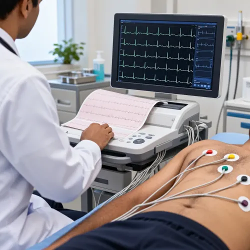
1. Expect the initial assessment to include blood pressure, pulse, temperature, oxygen saturation, and a physical examination. The clinician will also perform a 12-lead ECG for chest pain to record the heart’s electrical activity.
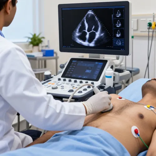
2. Keep the ECG vs echocardiogram distinction clear: an ECG shows electrical activity, while an echocardiogram uses ultrasound to examine heart structure, pumping function, and valves. The clinician chooses an echocardiogram when findings such as a murmur, breathlessness, an abnormal ECG, or suspected heart failure suggest a structural or functional problem.
3. If acute coronary syndrome is suspected, high-sensitivity troponin blood tests help detect heart-muscle injury. Clinicians interpret the result with your symptoms, symptom timing, ECG findings, the assay’s reference limit, and serial changes; one normal ECG or early troponin result does not always exclude danger.
The Heart Clinic - Dr. Satyajeet - 08048066834 in Pune can connect your symptom pattern and initial findings with appropriate testing instead of treating every episode as routine indigestion. Share any previous reports, diagnoses, allergies, and blood-thinner details before testing begins.
Related services
Coronary Angiogram A coronary angiogram is a diagnostic image, which uses dye and special X-rays to show the inside of your coronary (heart) arteries. View service → |
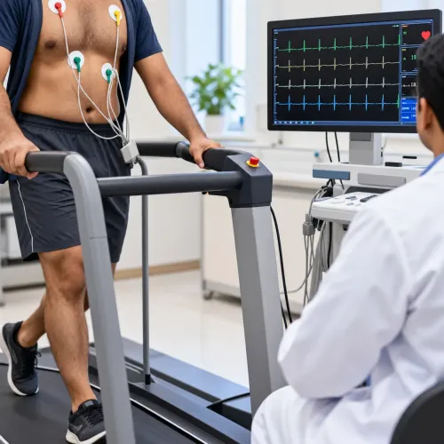
TMT (Stress Test) TMT (Stress Test) in Aundh, Baner, Pune TMT is the abbreviation for Treadmill Test. The TMT Testing takes into account, the measurement of the blood circulation inside the body (blood... View service → |
How clinicians choose further tests after the initial assessment
A structured chest pain risk assessment determines whether you need observation, repeat testing, coronary CT angiography, stress imaging, or another investigation. With stable chest pain and no known coronary disease, low-risk patients may not need immediate testing. Intermediate-risk patients often need coronary CT angiography or stress test selection based on the factors below.
| Option | Main advantage | What affects the choice |
|---|---|---|
| Observation or repeat ECG and troponin | Detects evolving heart-muscle injury or changing electrical findings | Used when symptoms, timing, examination, or initial results leave uncertainty |
| Exercise ECG | Tests symptoms and ECG changes during exertion | Requires adequate exercise capacity and an interpretable resting ECG |
| Coronary CT angiography | Shows plaque and narrowing, including non-obstructive disease | Kidney function, iodinated contrast concerns, radiation, coronary calcification, heart rhythm, and local expertise |
| Stress imaging | Assesses blood flow or heart function under stress | Useful when exercise ECG is unsuitable, baseline ECG changes limit interpretation, or earlier testing was inconclusive |
A normal early result does not end the assessment when symptoms continue or the clinical risk remains concerning. The work-up may instead target an arrhythmia, myocarditis, pericarditis, heart failure, valve disease, cardiomyopathy, or pulmonary and aortic causes; echocardiography helps assess structure, pumping function, and valves.
Chest pain after stent placement, bypass surgery, or known coronary disease needs reassessment, not self-treatment as indigestion.
Frequently asked questions
When does chest pain need emergency help immediately?
Call emergency services for pressure, squeezing, fullness, heaviness, or severe chest pain lasting more than a few minutes. Call again without waiting if it eases and returns.
How does heart-related chest pain differ from common non-cardiac pain?
Heart-related pain often feels like pressure, squeezing, fullness, heaviness, or severe discomfort. Non-cardiac pain can have different triggers and features, but new or unexplained chest pain still needs medical assessment.
Which chest-pain patterns and risk factors lower the threshold for evaluation?
Seek evaluation sooner for exertional or recurring pain, pain with breathlessness or sweating, and pain in someone with cardiovascular risk factors or known heart disease.
What happens during a first heart evaluation?
Clinicians review your symptoms and risk factors, examine you, and usually perform an ECG. They use those findings to decide whether blood tests, imaging, stress testing, or other tests are needed.
Related service
ECG ECG Treatments in Aundh, Baner, Pune Echocardiography is a test that uses sound waves to produce live images of your heart. The image is an echocardiogram. View service → |