

A resting 2D echocardiogram is recommended when symptoms, examination findings, medical history or another test raises a specific question about the heart’s structure or performance. By the end, you will know which findings commonly lead to a referral, what the report measures, when another cardiac test is more suitable and how to prepare for the appointment.
Key takeaways
- Ask about a resting 2D echo when symptoms or tests suggest structural heart disease.
- Ejection fraction estimates how much blood the left ventricle pumps per contraction.
- A 2D echo cannot replace stress testing, coronary imaging or rhythm monitoring.
- Bring medication details and earlier heart reports to improve result interpretation.
Which symptoms and findings justify a resting 2D echo?
A resting transthoracic 2D echo is appropriate when symptoms, examination findings, medical history or earlier tests raise a specific question about the heart’s structure or pumping function. It is not a general health check for an entirely asymptomatic person with a normal examination and no cardiovascular history.
- Unexplained breathlessness, reduced exercise tolerance or leg swelling can prompt evaluation for suspected heart failure, raised pressure in the lungs or valve disease.
- A new or changing heart murmur deserves an echo when it accompanies breathlessness, chest discomfort, fainting, an abnormal examination or other evidence of significant valve disease.
- An abnormal ECG, chest X-ray or blood-test result can provide the reason to look for enlarged chambers, impaired contraction, cardiomyopathy or pericardial disease.
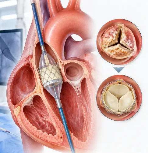
- Known valve disease, congenital heart disease, pulmonary hypertension, cardiomyopathy, a previous heart attack, or follow-up after cardiac treatment can require imaging to assess change.
| Finding or history | Clinical question for the echo |
|---|---|
| Abnormal ECG | Is there structural heart disease or impaired contraction? |
| Family history of cardiomyopathy | Are the chambers or heart muscle abnormal? |
| Prior heart attack | Is there lasting wall-motion or pumping impairment? |
| Known valve disease | Has narrowing or leakage changed? |
Pregnancy does not prevent a medically indicated echo because it uses ultrasound, not ionising radiation. A normal scan lowers concern for major structural disease at that time, but it does not exclude intermittent rhythm problems or every cause of breathlessness.
What does the echo report measure, and what does ejection fraction mean?
An echo report measures how the heart is built and how it works: chamber size, wall thickness, muscle movement, valve opening and leakage, right-heart function, filling patterns and estimated pulmonary artery pressure.
1. Start with LVEF. Ejection fraction is the percentage of blood the left ventricle ejects with each contraction. An LVEF of about 50% to 70% is commonly described as preserved ejection fraction, although the laboratory’s reference range matters.
When image quality allows, the biplane Simpson method is preferred to visual estimation because it calculates volume from traced ventricular discs.
2. Read the remaining measurements alongside LVEF. Regional wall-motion abnormalities can indicate previous or active heart-muscle injury. Valve gradients show pressure differences across narrowed valves, while regurgitation describes backward leakage. Chamber enlargement, increased wall thickness, impaired relaxation, right-ventricular weakness and raised estimated pulmonary artery pressure can reveal important disease even when LVEF is preserved.
3. Compare the result with symptoms, blood pressure, rhythm, examination findings and earlier scans. Heart-failure symptoms can occur with preserved ejection fraction when the ventricle is stiff, filling pressures are high, valves are abnormal or rhythm and lung disease limit circulation. This is called heart failure with preserved ejection fraction, or HFpEF.
A small LVEF change does not automatically prove recovery or deterioration. Different machines, readers and loading conditions can shift the measurement, so the clinical picture and scan quality determine how much weight to give one number.
When is a resting 2D echo not the right study?
A resting transthoracic 2D echo is the wrong study when the question concerns blood flow during exertion, a fleeting rhythm disturbance, or a structure that ultrasound cannot image clearly through the chest. It assesses chambers, valves, wall motion and pumping at rest; it does not directly show most coronary-artery blockages.
| Test | Main purpose | When it applies |
|---|---|---|
| Doppler echocardiography | Measures blood-flow speed and direction | Suspected valve narrowing, valve leakage or raised pressure |
| Stress echo | Compares wall motion at rest and during exercise or medicine-induced stress | Suspected inducible ischaemia or reduced exercise capacity |
| Transoesophageal echocardiography | Images structures from the oesophagus | Poor transthoracic views, prosthetic valves, clots in the left atrial appendage or suspected endocarditis |
| Contrast echo | Improves visibility of the inner ventricular border | Poor image quality or uncertain wall motion and ejection fraction |
Ask whether another test is needed when:
- Chest discomfort suggests coronary disease. Options include stress echo, CT coronary angiography or another ischaemia test selected for your risk and ability to exercise.
- Palpitations, fainting or dizziness occur unpredictably. A Holter monitor, event recorder or implantable loop recorder captures rhythm; a normal echo cannot exclude an intermittent arrhythmia.
- Breathlessness or chest pressure appears with activity but the resting scan is normal. Exercise testing or stress imaging can reveal a problem that rest conceals.
Persistent or worsening symptoms require reassessment rather than reliance on an old normal report.
What happens during a 2D echo, and how should you prepare?
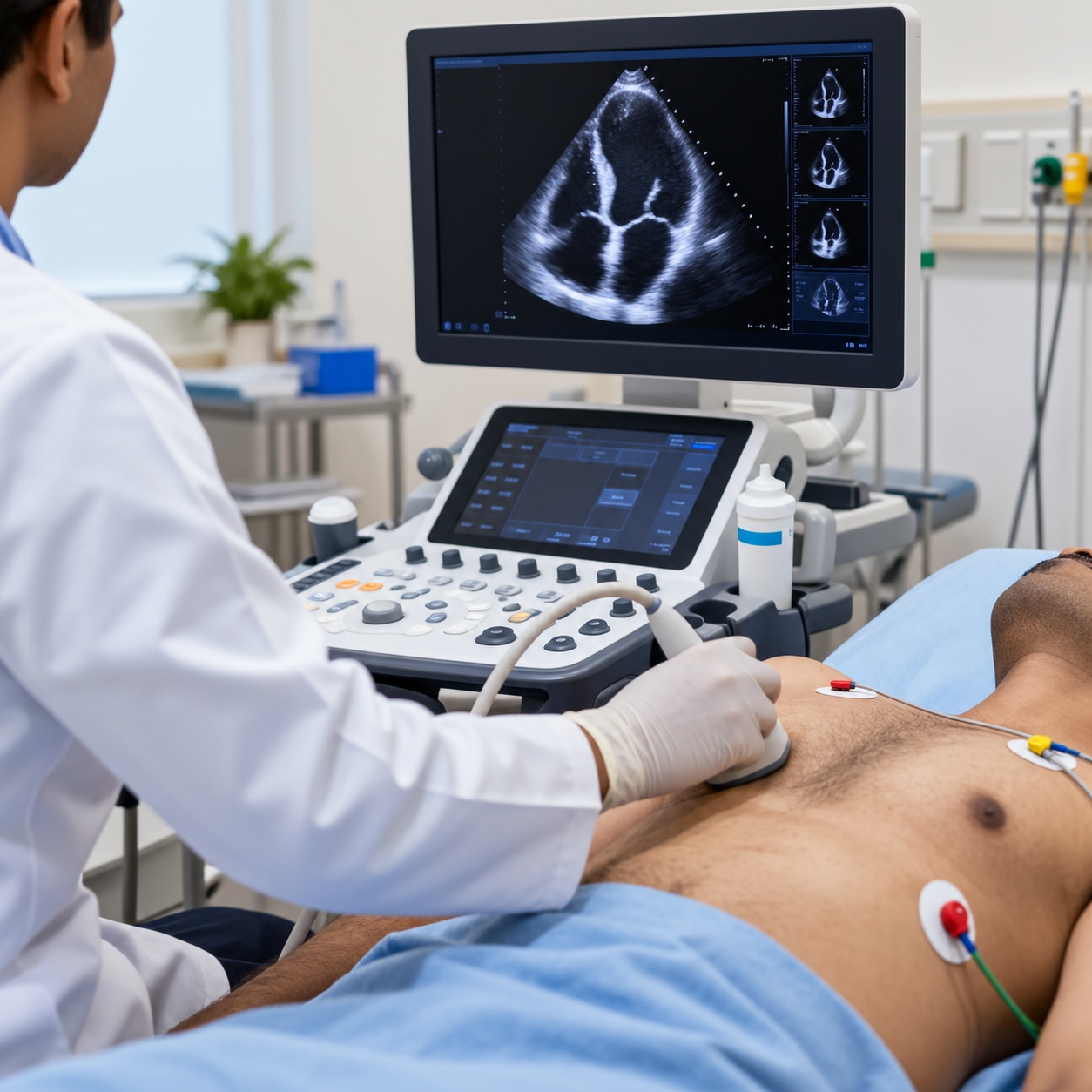
A standard resting 2D echo usually takes 20 to 45 minutes and does not require sedation. Wear a two-piece outfit that allows easy access to your chest; you may change into a gown.
- Lie on your back and then turn partly onto your left side. The sonographer places ECG leads on your chest to track your heartbeat while recording images.
- The sonographer applies clear gel and moves the probe over several locations, including the left side of your chest, the area below the breastbone and the upper abdomen. Firm pressure helps obtain clearer views.
- Hold your breath for a few seconds when instructed. This steadies the heart and diaphragm; you can breathe normally between recordings.
- You can eat and drink before an echocardiogram. Take your usual medicines unless the ordering clinician gives different instructions; fasting is not needed for a routine resting study.
- Bring your prior echo report, ECGs, discharge summaries, medication list and referral documents. Tell the team about a prosthetic valve, implanted device, previous heart surgery or endocarditis.
- You do not usually need an escort. Bring someone if you feel unsteady, have difficulty lying flat or need help understanding instructions.
Good 2D echo preparation includes asking whether the clinic needs earlier images for comparison. The Heart Clinic - Dr. Satyajeet - 08048066834 can use those records to help the cardiologist prioritise relevant views.
Related service
2D Echo 2D Echocardiography or 2D Echo of heart is a test in which ultrasound technique is used to take pictures of heart. View service → |
How should a normal, abnormal or limited result guide follow-up?
A result does not set follow-up by itself. Interpret it alongside the clinical picture, because a normal echocardiogram can coexist with intermittent rhythm disease, early heart failure with preserved ejection fraction, or coronary narrowing that appears only with exertion. Read the report with:
- current symptoms
- blood pressure during evaluation
- heart rhythm and ECG findings
- physical examination, including a murmur or congestion
- earlier echocardiograms and relevant tests
An abnormal result needs follow-up matched to the finding and its change over time. In established valve disease, repeat imaging follows lesion severity and changes in symptoms or examination, rather than an automatic annual schedule. A rising pulmonary artery pressure or new wall-motion abnormalities can change urgency.
A limited study means the images did not answer the question reliably, not that the heart is abnormal. Body habitus, lung disease or positioning can hide endocardial borders; ask whether to repeat imaging, use ultrasound-enhancing contrast or choose another study.
During pregnancy, a medically indicated transthoracic echo remains appropriate because it uses no ionising radiation; symptoms and suspected cardiac disease determine timing.
Frequently asked questions
Which symptoms and findings justify a resting 2D echo?
Breathlessness, chest discomfort, swelling, fainting, a heart murmur, abnormal ECG, known heart disease or suspected heart failure can justify a resting 2D echo when a clinician needs to assess structure or pumping function.
What does ejection fraction mean on an echo report?
Ejection fraction is the percentage of blood the left ventricle pumps out with each contraction. It helps assess systolic pumping function but does not describe every aspect of heart health.
When is a resting 2D echo not the right study?
A resting echo does not show every cause of chest pain or palpitations. Stress echocardiography, an ECG, Holter monitoring, coronary CT angiography or cardiac MRI may answer a different clinical question.
How should you prepare for a 2D echo and follow up afterward?
A standard resting transthoracic echo usually needs no fasting. Wear comfortable clothing, bring previous reports and medicines, and ask the ordering clinician to explain whether the result is normal, abnormal or limited.