Valve narrowing, leakage and weak or stiff pumping can produce similar symptoms, yet each problem leaves different clues in an echocardiogram. By the end, you will know which measurements matter, how to prepare, what the test can and cannot answer, and how to judge whether a report is complete.
Key takeaways
- Doppler measures blood-flow speed and pressure gradients across narrowed or leaking valves.
- Ejection fraction estimates how much blood the left ventricle pumps with each beat.
- Wear a two-piece outfit and bring previous cardiac reports to your appointment.
- Match the test to the clinical question before choosing a 2D Echo provider.
How a 2D Echo Reveals Valve Narrowing, Leakage and Abnormal Pumping
Valve narrowing appears when leaflets become thickened, calcified, or restricted and open less than normal. Two-dimensional imaging shows the valve’s shape and movement; colour Doppler shows turbulent flow through the narrowed opening, while spectral Doppler measures velocity and the pressure gradient across it. This helps assess aortic, mitral, tricuspid, and pulmonary stenosis.
Leakage, or regurgitation, occurs when a valve fails to close properly. Colour Doppler can show the backward jet, while 2D images may reveal prolapse, flail leaflets, rheumatic deformity, infection-related damage, or enlargement of the valve ring. A complete 2D Echo Test in Pune should therefore include Doppler measurements, not only still images or moving pictures.
A standard transthoracic study can also identify these pumping and structural abnormalities:
- Reduced left-ventricular ejection fraction, indicating weaker squeezing
- Regional wall-motion abnormality, where one area contracts less or not at all
- Enlarged left or right ventricle, atrium, or both
- Thickened ventricular walls, as seen with long-standing pressure overload
- Impaired relaxation or raised filling pressure, even when ejection fraction is normal
- Pericardial effusion, congenital structural defects, and pressure-related right-heart changes
M-mode and 2D measurements assess chamber size, wall thickness, and fractional shortening. Tissue Doppler evaluates myocardial motion, complementing mitral inflow, left-atrial size, and tricuspid-regurgitation velocity when assessing diastolic function. The test does not by itself diagnose every cause of chest pain, intermittent rhythm disturbance, or exertional breathlessness.
Reading the Numbers: Ejection Fraction, Doppler Flow and Chamber Measurements
A valve is not graded from one Doppler number. For aortic stenosis, interpret peak velocity, mean pressure gradient and calculated valve area together. The gradient depends on blood flow: low flow from weak ventricular contraction, or high flow from fever, anaemia or another high-output state, can produce apparently conflicting results.
Left-ventricular ejection fraction (LVEF) is the percentage of blood pumped out between filling and contraction, calculated from end-diastolic and end-systolic volumes, commonly by Simpson’s biplane method. Broad categories are reduced below 40%, mildly reduced at 41–49%, and preserved at 50% or higher.
Fractional shortening uses a change in ventricular diameter, so it is not interchangeable with LVEF.
A normal LVEF does not exclude a pumping problem. Filling and relaxation are assessed from several findings together:
- Mitral inflow patterns
- Tissue-Doppler e′ velocity
- Tricuspid-regurgitation velocity
- Left-atrial volume
For valve leakage, colour-jet area alone can mislead because machine settings, jet direction and loading conditions alter its appearance. The report may combine vena contracta, effective regurgitant orifice area, regurgitant volume, pulmonary-vein flow and chamber remodelling.
Check chamber size, wall thickness, right-heart measurements and pulmonary pressure alongside the labels mild, moderate or severe. Blood pressure, rhythm, symptoms and earlier studies can change interpretation. A complete 2D echo includes colour, spectral and tissue Doppler—not only still images.
What Happens During the Appointment and How to Prepare
For an uncomplicated transthoracic 2D echo, preparation is minimal: arrive in comfortable clothing, bring your referral and previous cardiac reports, and take prescribed medicines unless the clinic gives different instructions. Do not assume fasting or medication changes are needed.
1. Before the scan, confirm which examination was ordered. A routine TTE differs from a stress echo, contrast echo and transesophageal echo (TEE). Ask whether the appointment includes colour Doppler, spectral Doppler and tissue Doppler, not only moving images.
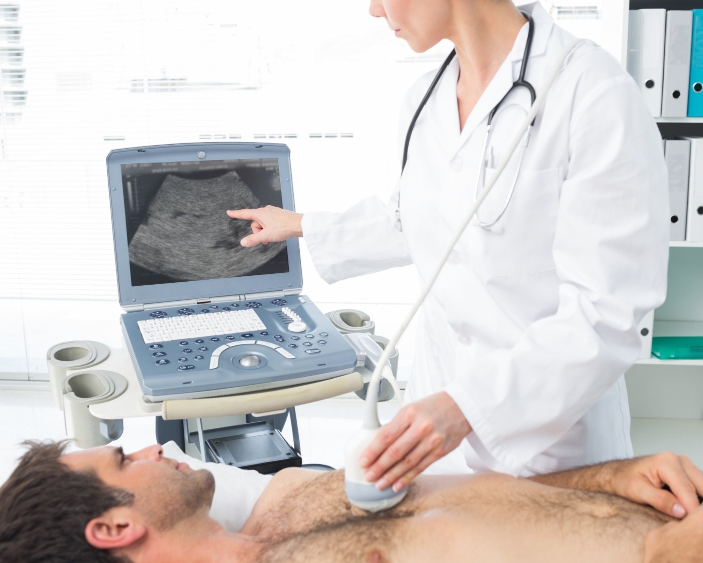
2. During the scan, you will remove clothing from the upper body and lie on your back or partly on your left side. Staff attach small ECG electrodes, apply gel to your chest and move a handheld transducer across several positions.
3. The sonographer may ask you to turn, hold your breath briefly or breathe normally. Transducer pressure can feel uncomfortable over the ribs, but the test does not use needles, radiation or incisions. A routine study often takes 20 to 45 minutes.
4. Immediately afterward, staff wipe off the gel and remove the electrodes. You can dress, eat, drive and return to normal activities unless another procedure is scheduled; routine TTE does not require sedation or an escort.
The sonographer may not explain every finding during the scan. The cardiologist reviews the images and Doppler measurements before issuing the report, so ask when and how you will receive the final interpretation.
Choosing the Right Cardiac Test for the Clinical Question
A routine transthoracic 2D echo is appropriate when the question concerns heart structure and function at rest: valve narrowing or leakage, chamber size, wall motion, pumping strength, pericardial fluid, or many congenital abnormalities. It cannot explain every chest-pain episode, detect every intermittent rhythm problem, or show coronary blockage reliably.
Choose the protocol for the clinical question, not the cheapest examination labelled “2D echo.”
| Test | Best information | When it applies |
|---|---|---|
| ECG | Electrical activity, conduction and rhythm at that moment | Palpitations, chest pain, fainting, or suspected heart attack |
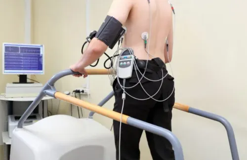
| TMT | Heart-rate and ECG response during exercise | Exercise-related symptoms when the person can walk and an exercise ECG is suitable |
| Stress echo | Wall motion and pumping response during exertion or medicine-induced stress | Suspected inducible ischaemia or exercise-related valve and pumping assessment |
| TEE | Close views of valves, prosthetic valves, left atrial appendage, clots and infection | When transthoracic images are inadequate or these structures need detailed assessment; it involves an oesophageal probe and sedation planning |
| CT | Coronary anatomy, calcium, aorta and selected structural detail | Suspected coronary disease or aortic and congenital anatomy questions |
| MRI | Precise volumes, blood flow, scar, inflammation and tissue characterisation | Cardiomyopathy, myocarditis or unclear ventricular findings |
| Rhythm monitoring | Heart rhythm over hours to weeks | Palpitations, dizziness or fainting that an ECG misses |
Ask whether the facility can arrange contrast echo, stress echo or TEE when the initial study cannot answer the referral question.
How to Compare Reports and Choose a 2D Echo Provider in Pune
A reliable report gives you more than “normal” or “mild regurgitation.” Check that it identifies the valve examined, includes Doppler findings, lists chamber dimensions, wall thickness, ejection fraction, right-ventricular function and estimated pulmonary pressure, and states the rhythm and blood pressure when recorded.
| Report item | What to check | Why it matters |
|---|---|---|
| Valve assessment | Valve area, peak velocity, mean gradient and regurgitation grade | Separates narrowing from leakage and supports severity grading |
| Pump function | Ejection fraction, wall-motion description and chamber size | Shows whether contraction is reduced, regional or preserved |
| Filling and pressure | Tissue Doppler, left-atrial size and pulmonary-pressure estimate | Helps explain breathlessness and impaired relaxation |
| Comparison | Date-linked measurements and access to earlier images | Reveals a real change better than comparing “mild” or “moderate” alone |
Ask whether the study is a complete transthoracic examination with colour, spectral and tissue Doppler, not a technician-produced printout of still images. A qualified 2D Echo Specialist in Pune should interpret the images alongside your symptoms, blood pressure, rhythm, medicines and previous scans.
Before booking 2D Echo Services in Pune, confirm who signs the report and whether the centre can arrange contrast echo, stress echo or TEE when the first study cannot answer the referral question. TEE is semi-invasive and involves sedation planning, so it is not a routine upgrade.
The Heart Clinic - Dr. Satyajeet - 08048066834 can be considered when you need specialist interpretation and continuity between reports rather than a standalone measurement sheet.
Related service
2D Echo 2D Echocardiography or 2D Echo of heart is a test in which ultrasound technique is used to take pictures of heart. View service → |
Frequently asked questions
What can a 2D Echo reveal about heart valves?
It shows valve shape and movement, while colour and spectral Doppler identify turbulent flow, velocity and pressure gradients linked to narrowing or leakage.
What does ejection fraction mean on a 2D Echo report?
Ejection fraction estimates the percentage of blood the left ventricle pumps out with each contraction and helps assess pumping function.
How should you prepare for a 2D Echo appointment?
Wear clothing that allows access to your chest, bring previous reports and medication details, and follow any instructions from the testing centre.
How do you choose between cardiac tests?
Choose the test that answers the clinical question: a resting 2D Echo assesses structure and pumping, while other tests evaluate rhythm, exercise response or coronary blood flow.
Keywords