

An ECG can show how electrical signals travel through your heart at the moment of recording, but its lines and intervals are easy to misread without context. You will learn what the main parts of a 12-lead tracing mean, which conditions it can suggest, and when a different or longer test is needed.
Key takeaways
- A standard 12-lead ECG uses 10 electrodes to create 12 electrical views.
- Clinicians assess waves, intervals, heart rate, rhythm, and conduction patterns.
- Use a Holter monitor for frequent symptoms and an event monitor for less predictable episodes.
- Seek urgent help for chest pain, fainting, severe breathlessness, or a sustained racing heartbeat.
What happens during a standard 12-lead ECG
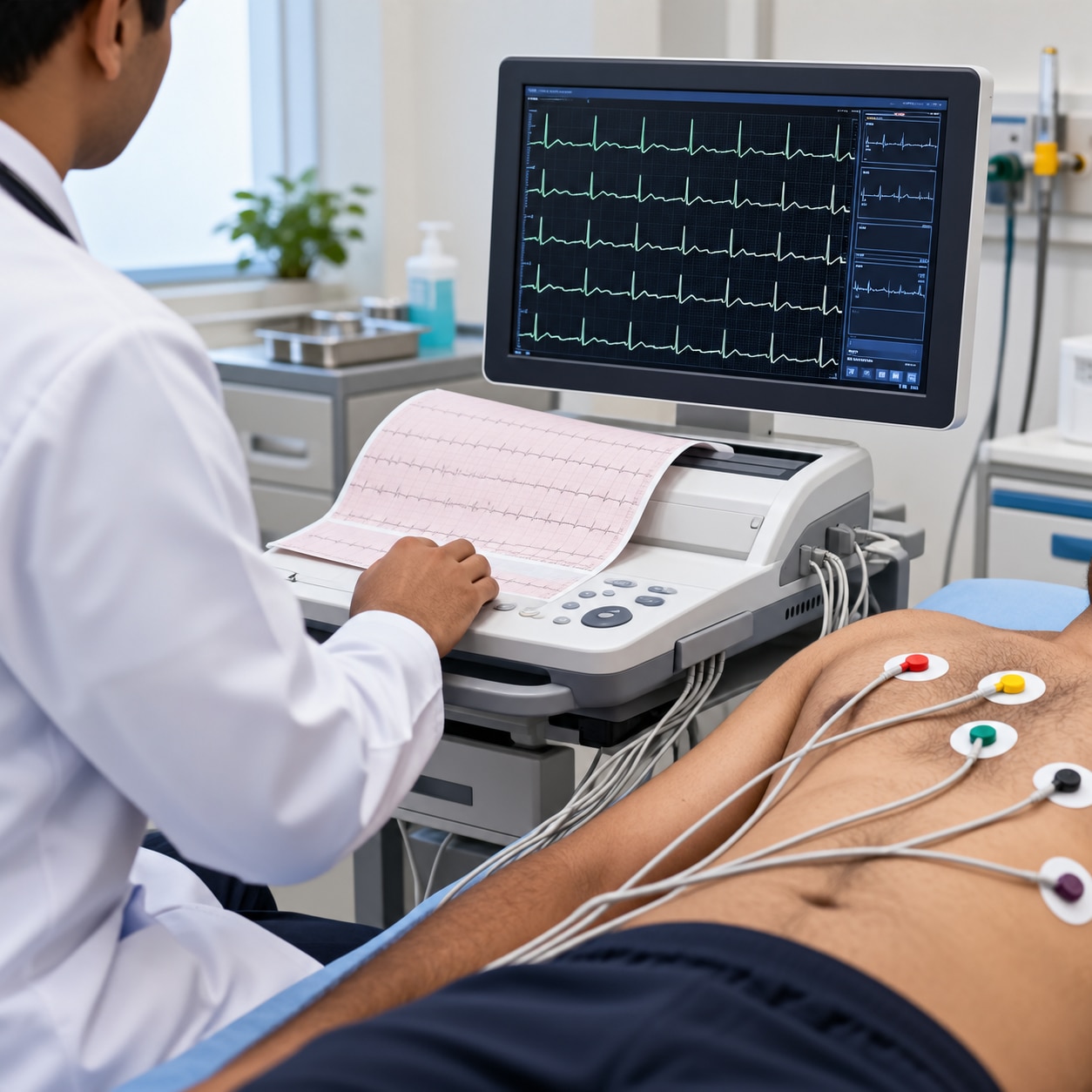
A 12-lead ECG records the heart’s electrical activity through 10 skin electrodes: four on the limbs and six across the chest. These electrodes generate 12 electrical views while you lie still, usually for only a few seconds.
Resting ECG preparation means exposing the electrode sites, cleaning the skin, and removing excess oil or sweat so the sensors adhere firmly.
Accuracy suffers when the tracing picks up muscle or electrical interference. Common problems include:
- Moving, talking, coughing, or shivering during recording
- Loose electrodes or poor skin contact from sweat, hair, lotion, or dry skin
- Incorrect electrode placement, which can mimic a heart attack, shift the electrical axis, or resemble dextrocardia
The ECG is a short snapshot, not continuous monitoring. A normal tracing between episodes does not exclude an intermittent arrhythmia; a Holter or event monitor may be needed when symptoms come and go. Standard leads also do not show every heart region equally well, so suspected right-ventricular or posterior infarction can require additional leads.
If an ECG conflicts with your symptoms or an earlier tracing, ask whether electrode placement was checked and the test repeated. Computer interpretations are screening aids; a clinician must review the tracing alongside your symptoms, examination, and previous ECGs.
How clinicians read the waves, intervals, and rhythm
Clinicians read an ECG as a sequence: atrial activation, ventricular activation, recovery, rate, and regularity.
- P wave: atrial electrical activation; its shape and presence help identify sinus rhythm.
- PR interval 120–200 milliseconds: conduction from atria through the atrioventricular node to the ventricles.
- QRS complex: ventricular activation; a wide QRS 120 milliseconds or more indicates delayed ventricular conduction.
- ST segment: the early recovery phase; ST elevation or ST depression can support myocardial ischaemia or infarction.
- T wave: ventricular recovery; T-wave changes can support myocardial ischaemia or infarction.
- Rate: the number of ventricular beats per minute.
- Rhythm regularity: whether beats occur at consistent intervals and whether atrial and ventricular activity stay linked.
Hyperkalaemia can progress from tall peaked T waves to PR prolongation, P-wave loss, QRS widening, and a sine-wave pattern, but a blood potassium test remains necessary.
Symptoms, serial ECGs, and other tests determine the significance of ST and T-wave findings. A single normal ECG does not reliably exclude acute coronary syndrome.
Standard leads do not display every myocardial region equally, so suspected right-ventricular or posterior infarction may require additional leads. Incorrect placement can imitate infarction, axis deviation, or dextrocardia; repeat the ECG after checking placement.
Computer interpretations are screening aids, not final diagnoses: a clinician must review the tracing with symptoms, examination, medicines, previous ECGs, and laboratory results.
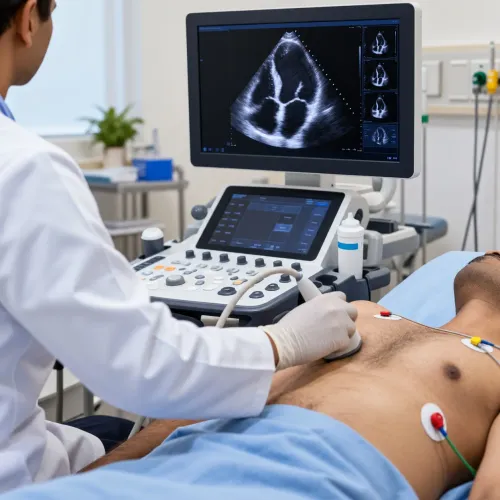
An ECG cannot show blocked arteries, valve structure, pumping strength, or every structural problem. Echocardiography assesses these features; TMT or other stress testing evaluates exertional changes, and coronary imaging may be needed. Blood tests can identify anaemia, thyroid disease, and electrolyte imbalance.
Which rhythm test fits the timing of your symptoms?
Match the test to how often your symptoms occur. A resting ECG fits symptoms happening now, such as ongoing palpitations, a fast pulse, dizziness, or fainting. It records only a few seconds, so a normal result between episodes does not exclude intermittent atrial fibrillation or another rhythm problem.
| Test | When it fits | What it can capture |
|---|---|---|
| Resting ECG | Symptoms are present in the clinic, or a baseline is needed | Current rate, rhythm, conduction, and ST-T changes |
| Repeat ECG | Symptoms return, the first tracing was unclear, or your condition changes | A new rhythm compared with the earlier tracing |
| 24–48-hour Holter monitor | Symptoms occur daily or every few days | Continuous rhythm during ordinary activity and sleep |
| Event monitor | Episodes are less frequent and you can activate the device when symptoms start | The rhythm during a reported palpitation, dizzy spell, or near-faint |
| Patch monitoring | Symptoms occur weekly or less predictably | Continuous recording over a longer wearable-monitoring period |
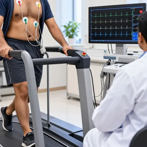
| Exercise ECG | Symptoms begin with walking, climbing, or other exertion | Rhythm and ECG changes during increasing workload |
| EP study | Recurrent or dangerous arrhythmia remains unexplained after non-invasive testing | Catheter-based mapping that can locate and sometimes trigger the abnormal circuit |
Tell the clinician how often episodes happen, how long they last, and what triggers them. Seek urgent assessment for fainting with palpitations, severe breathlessness, or chest pressure; do not wait for an outpatient monitor.
What to do after the ECG and when to seek help
Ask for two things before leaving: the printed ECG tracing and a clinician’s written rhythm interpretation. The report should include the heart rate, rhythm, PR interval, QRS duration, QT/QTc, and any ST-segment or T-wave changes. Treat the computer-generated conclusion as a screening aid, not a diagnosis.
| Result | What it means | Follow-up |
|---|---|---|
| Clinician-confirmed normal ECG | No abnormality appears during recording | Continue the recommended plan; a normal tracing does not exclude intermittent atrial fibrillation or episodic tachycardia |
| Abnormal rhythm or conduction finding | The tracing needs clinical context | Ask whether you need a repeat ECG, 24–48-hour Holter monitor, event monitor, or longer patch monitor |
| ST-segment or T-wave change | Possible ischaemia or infarction, not a diagnosis by itself | Seek prompt assessment and ask whether serial ECGs and blood tests are needed |
If palpitations, fainting, or rapid heartbeat occur between appointments, record the time, duration, activity, and symptoms. At The Heart Clinic - Dr. Satyajeet - 08048066834, a clinician can use that pattern to choose monitoring rather than treating one normal ECG as conclusive.
Call emergency services immediately for:
- Chest pain, pressure, or tightness lasting more than a few minutes
- Fainting, severe breathlessness, or a sustained racing heartbeat with dizziness
- New weakness on one side, facial drooping, trouble speaking, or confusion
A normal ECG does not safely rule out acute coronary syndrome when chest pain is present.
Related services
Electrophysiological Study (EP study) Electrophysiological Study (EP study) An electrophysiological study (EP study) is a test used to evaluate your heart’s electrical system and to check for abnormal heart rhythms. View service → |
ECG ECG Treatments in Aundh, Baner, Pune Echocardiography is a test that uses sound waves to produce live images of your heart. The image is an echocardiogram. View service → |
Frequently asked questions
What happens during a standard 12-lead ECG?
You lie still while four limb electrodes and six chest electrodes record your heart’s electrical activity for a few seconds.
How do clinicians read ECG waves and intervals?
They examine the P wave, QRS complex, T wave, heart rate, rhythm regularity, and intervals such as PR and QT.
Which rhythm test fits the timing of symptoms?
A standard ECG suits symptoms happening during the appointment; a Holter monitor suits frequent episodes, while an event monitor suits less predictable symptoms.
When should you seek urgent help after an ECG?
Seek urgent care for chest pain, fainting, severe breathlessness, or a sustained racing or irregular heartbeat.
Related services
Holter Monitor If your heart seems to skip a beat, race, or work a bit too slow, you could have a condition known as arrhythmia. View service → |
Arrhythmia Arrhythmia An arrhythmia describes an irregular heartbeat – the heart may beat too fast, too slowly, too early, or irregularly. |