A resting ECG can reveal rate, rhythm, conduction timing and electrical patterns, but its brief recording can miss symptoms that come and go. You will learn how to prepare, read the main report terms, recognise misleading results, choose follow-up testing and compare local testing options safely.
Key takeaways
- Avoid creams and lotions before electrode placement.
- A normal resting ECG cannot rule out every heart problem.
- Ask what each rhythm term means before assuming it is dangerous.
- Choose Holter monitoring when symptoms do not occur during a clinic ECG.
What a Resting ECG Records and How to Prepare for It
A 12-lead ECG measures the heart’s electrical activity through electrodes on your skin. It does not measure blood flow, valve movement, pumping strength, or provide a direct image of the heart.
Before the test:
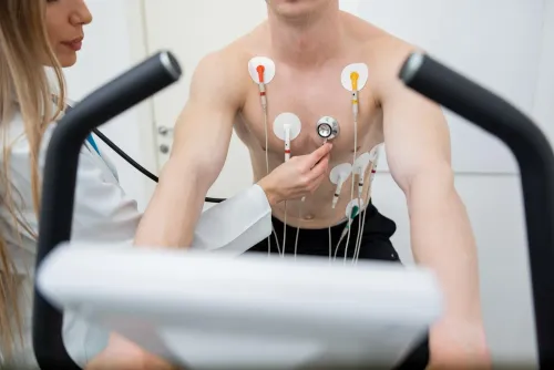
- Take your usual medicines unless the ordering clinician gives different instructions. Wear loose clothing that allows access to your chest, wrists and ankles.
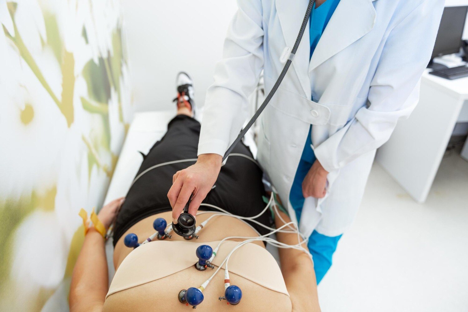
- Remove lotions, oils and creams from those areas. Staff will clean your skin, then place adhesive electrodes on your chest and limbs.
- Lie still and breathe normally while the machine records. Setup takes several minutes; the actual tracing usually lasts about 10 seconds.
- Expect no pain. An ECG sends no electricity into your body; the electrodes only detect signals.
- Give staff your correct name, age or date of birth, and other requested details before recording.
Movement, shivering, muscle tremor, oily skin, poor contact, loose leads and nearby electrical interference can create artefact that resembles a rhythm or ST-segment problem. Staff may repeat the tracing after improving contact and reducing movement. Incorrect electrode placement also matters: V1 or V2 placed too high can create false P-wave, Q-wave or T-wave changes.
An ECG is a sensible first test for palpitations, an irregular pulse, chest symptoms or suspected conduction problems. Choose an echocardiogram when the question concerns valve structure, heart chambers or pumping function. Searching for an ecg test near aundh helps locate testing, not confirm interpretation quality.
How to Read the Main ECG Lines and Common Rhythm Terms
1. The P wave shows atrial activation, when the upper chambers contract. The PR interval measures the signal’s travel from atria through the AV node to the ventricles; a prolonged or dropped PR-related beat can indicate heart block.
2. The QRS complex shows ventricular activation. A narrow complex usually follows the normal conduction route, while a premature ventricular contraction appears early with a wide, abnormal QRS, often followed by a pause.
3. The ST segment is the early recovery phase after ventricular activation. Elevation or depression can suggest injury or reduced blood flow, but movement, loose electrodes and electrical interference can imitate ST changes.
4. The T wave shows ventricular recovery. Heart rate counts beats per minute; rhythm regularity describes whether the intervals between beats are even. Electrical axis describes the overall direction of ventricular activation.
5. Sinus rhythm begins in the sinoatrial node. Sinus tachycardia is sinus rhythm above the usual resting range, not automatically an arrhythmia: fever, pain, anxiety, dehydration, anaemia, thyroid disease, stimulants and medicines can accelerate it.
Sinus bradycardia is slower sinus rhythm; sleep and athletic conditioning can cause it, while fainting, dizziness or poor exercise tolerance needs assessment.
6. Atrial fibrillation is an ECG diagnosis showing disorganised atrial activity and an irregular ventricular rhythm. Confirm it with a 12-lead ECG, not a pulse, symptom or smartwatch alert; intermittent episodes need ambulatory monitoring.
7. QT includes ventricular activation and recovery. QTc adjusts it for heart rate; Bazett correction can overcorrect at high rates and undercorrect at low rates. A prolonged QTc can increase dangerous rhythm risk, while a normal tracing does not exclude every problem.
When an Abnormal or Normal Result Needs Clinical Context
An ECG finding is significant when it matches your symptoms, examination and medical history, persists on a clean repeat tracing, or differs from an earlier ECG. A fast rate alone is not automatically an arrhythmia, and a slow rate can be normal during sleep or athletic training.
Clinicians interpret the tracing alongside your age, symptoms, medicines, electrolyte levels such as potassium and magnesium, physical examination and previous ECGs. Movement, muscle tremor, loose electrodes, baseline wander and electrical interference can imitate rhythm or ST-segment changes; improving contact and repeating the recording may resolve the apparent abnormality.
A computer-generated statement is an aid, not a diagnosis. Automated systems can misclassify atrial fibrillation, ischaemia and conduction problems, so a qualified clinician must review an abnormal result or one that conflicts with how you feel.
Keep a symptom timeline for the reviewing clinician:
- Record the exact start time, activity, duration, pulse regularity and heart rate if measured.
- Note palpitations, dizziness, fainting, chest pain, breathlessness and what you were doing when each began.
- Write down every medicine, stimulant or dose taken before the episode, including missed doses.
A normal 10-second ECG does not exclude an intermittent rhythm, early coronary disease or symptoms that occurred outside the recording. Chest pressure, severe breathlessness, fainting, sustained rapid heartbeat with weakness or near-fainting, or new neurological symptoms require urgent care, not a routine booking.
Choosing the Next Test When a Single ECG Is Not Enough
A normal clinic ECG is only a short snapshot, often about 10 seconds. If an intermittent episode is absent during recording, the result cannot reliably exclude it; suspected intermittent atrial fibrillation needs ambulatory monitoring even when the 12-lead ECG is normal.
Confirm atrial fibrillation with an ECG, not an irregular pulse, symptoms or smartwatch alert alone.
| Test | Best match | What it assesses |
|---|---|---|
| 24-hour Holter monitor | Symptoms occurring daily or almost daily | Rhythm during ordinary activities; a full day improves the chance of matching symptoms to beats |
| Event or patch monitor | Episodes occurring less often than daily | Rhythm over several days or weeks; longer duration improves capture |
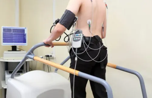
| TMT | Palpitations, chest discomfort or breathlessness triggered by exertion | ECG and blood-pressure responses while exercising |
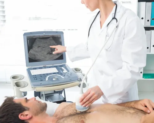
| Echocardiogram | Suspected structural or pumping problem | Heart chambers, valves and ventricular pumping function |
| Electrophysiological study | Recurrent or high-risk rhythm problems needing precise diagnosis or treatment planning | Detailed electrical mapping; it is invasive |
Choose a TMT when exertion reliably brings on symptoms and the clinician needs to observe exercise-related ECG or blood-pressure changes. Choose an echocardiogram when an ECG cannot answer questions about valves, chamber size or pumping strength. An electrophysiological study is considered when non-invasive tests do not define the rhythm or a procedure may be needed.
- Medication names and doses
- Previous ECGs and Holter reports
- Discharge summaries
- A symptom timeline with times, activities, duration, pulse, dizziness, fainting, chest pain and breathlessness
How to Compare ECG Testing Options in Aundh, Baner and Pune
The ecg test cost in aundh baner in pune has no single standard price. Compare what each quote includes, not just the headline fee.
| Option | Confirm before booking | Why it matters |
|---|---|---|
| Clinic recording | Recording, trained technician, cardiologist interpretation, consultation, and printed or digital report | A low fee may cover recording only |
| Fast or home service | Urgent reporting, home testing, travel charges, and stated turnaround time | Convenience can add separate charges |
| Repeat or follow-up | Correction of incorrect lead placement, repeat recording, and follow-up review | You should not face an unexpected repeat fee |
When comparing an ecg clinic in baner, ask these questions:
- Who places the leads, and what training do they have?
- Does a qualified clinician review an abnormal trace, rather than relying only on the computer statement?
- When will you receive the report?
- Can the site arrange Holter monitoring, echocardiography, or urgent escalation if the ECG does not explain your symptoms?
Searching for an ecg test near aundh improves access but does not prove clinical review. If chest pain, severe breathlessness, fainting, or sustained rapid palpitations with weakness or near-fainting is happening now, use urgent medical services instead of booking routinely.
For Baner patients seeking testing, interpretation, and follow-up decisions in one setting, The Heart Clinic - Dr. Satyajeet - 08048066834 is one local option to ask about these inclusions before booking.
Related services
ECG ECG Treatments in Aundh, Baner, Pune Echocardiography is a test that uses sound waves to produce live images of your heart. The image is an echocardiogram. View service → |
Arrhythmia Arrhythmia An arrhythmia describes an irregular heartbeat – the heart may beat too fast, too slowly, too early, or irregularly. |
Frequently asked questions
What does a resting 12-lead ECG record?
It records the heart’s electrical activity through skin electrodes. It does not directly show blood flow, valve movement, or pumping strength.
How should you prepare for an ECG test?
Wear clothing that allows chest access, avoid applying creams or lotions beforehand, and tell the technician about symptoms and medicines.
Does an abnormal ECG always mean heart disease?
No. Results need clinical context, including symptoms, medical history, medicines, and the specific ECG finding.
What test is used when a single ECG misses symptoms?
A Holter monitor records continuously for a longer period, while an event monitor records during intermittent symptoms.
Related services
Holter Monitor If your heart seems to skip a beat, race, or work a bit too slow, you could have a condition known as arrhythmia. View service → |
Electrophysiological Study (EP study) Electrophysiological Study (EP study) An electrophysiological study (EP study) is a test used to evaluate your heart’s electrical system and to check for abnormal heart rhythms. View service → |
Keywords